New equipment
We sell only in Africa
Free delivery Abidjan
ref : 10619
Year of mfg. : 2026
Available
We sell only in Africa
Free delivery Abidjan
New equipment
Optional OCTA module (contact us)
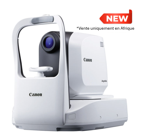
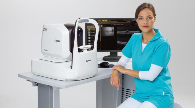
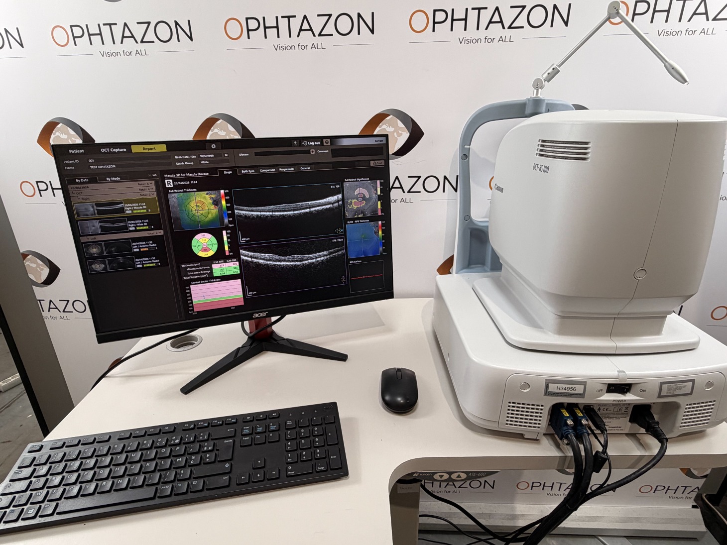
Xephilio OCT-A1 the latest Canon Medical High Resolution 3um optical coherence tomography for outstanding performance and exceptional ease of use you can rely on every day. High definition imaging quality and a host of automated features optimize and simplify your examinations with fast acquisition times while increasing your patients efficiency and comfort.
High Resolution OCT
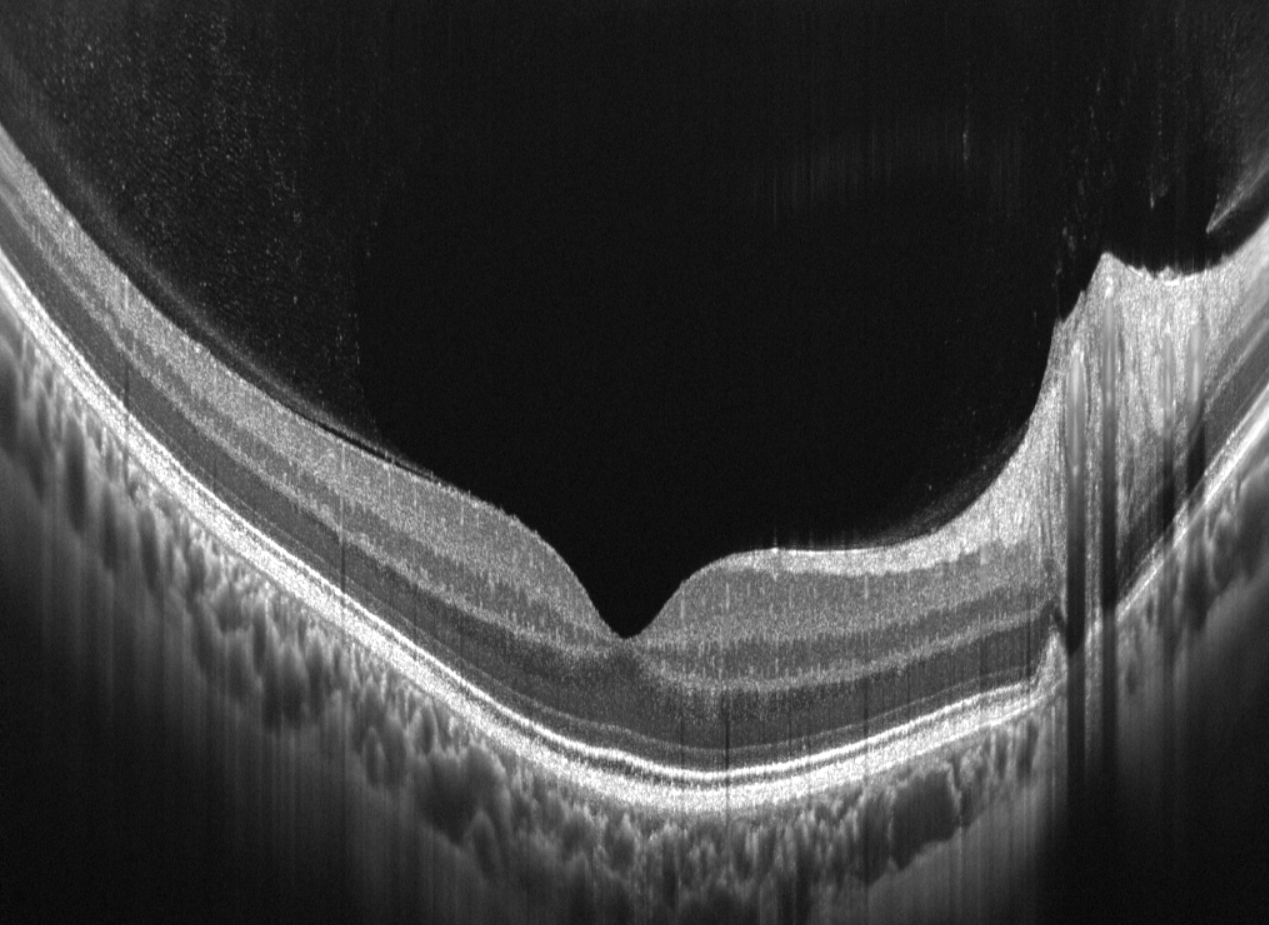
Xephilio OCT-A1 offers superb image quality. The 3 micron optical resolution ( 1.6 μm digital) enables excellent differentiation of structures and individual layers of the retina.
A complete exam with just 3 clicks
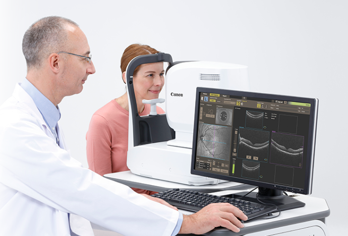
Examinations with the Xephilio OCT-A1 are extremely simple and therefore easy to delegate. With just 3 mouse clicks, scans be acquired. A complete range of intelligent functions enables fully automated examinations. In addition the auto-re-scan function intervenes if a patient makes unwanted eye movements and automatically compensates for any motion artefacts.
Automated anterior tracking
After clicking with the mouse on just roughly the center of the pupil, the OCT-A1 's automated anterior tracking will take over and will automatically detect and maintain the exact center, even when the patient is making eye movements or blinking.
Automatic image optimization
A second mouse click will initiate the scanning and the device will automatically optimize the focus and coherence gate for the highest signal quality and the best possible examination results. After confirmation of the pre-view scan images, the third click will start the actual acquisition which only takes seconds.
Real-time retinal tracking
The OCT-A1 is equipped with a Scanning Laser Ophthalmoscope. This SLO system allows for real-time retinal tracking. By detecting and compensating movements, the impact of small involuntary movements that could cause motion artefacts is greatly reduced and image quality is greatly improved.
High Image resolution
With Xephilio OCT-A1, up to 200 scans can be averaged to achieve an image resolution that allows you to see in detail both the layer structure as well as the vitreous pleated structure. The device combines high resolution image with a particularly wide scan width of up to 13 mm!
Enhanced depth visualization
For optimal imaging, the system offers special scan modes for both vitreous and choroid imaging.
Reliable 10 layer recognition
Canon’s Xephilio OCT-A1 can automatically detect and distinguish 10 layers of the retina – including Bruch’s membrane (BM).
Anterior imaging
With the optional Anterior Segment Adapter ASA-1, the 3 micron resolution of Xephilio OCT-A1 also gives you the ability to image the anterior segment of the eye in great resolution. The width of Anterior scans can be 6 or 9 mm.
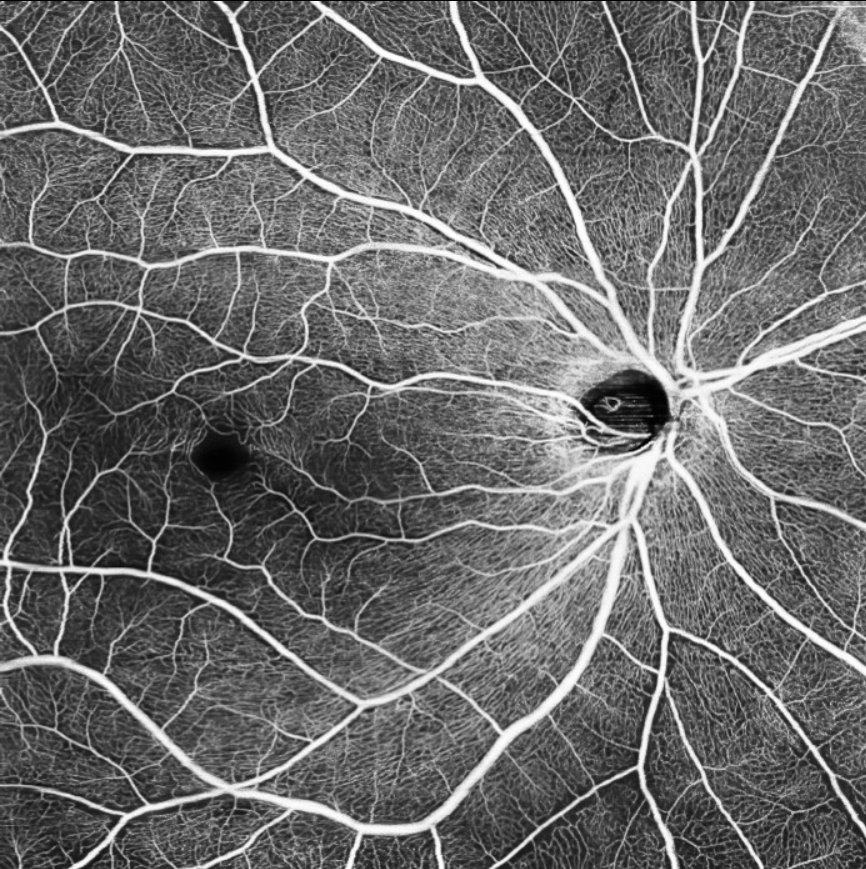
OCT Angiography
With the optional Angio Expert software module, the OCT-A1 can provide you with amazing OCTA images, showing the great optical qualities of the device.
3D Visualization
The unique optical characterics of the OCT-A1 also provide a very strong 3D visualization. The 3D images can support the clinical diagnosis and could be used for patient education as well.
Fast and precise follow-up
The SLO also enables accurate follow-up examinations by automatically adjusting to the same scan position as used in the previous exam. The SLO image of the center part of the retina is compared with the previous study: like a digital finger print for reliable comparison, the software automatically selects identical scan parameters.
Angiographie OCT
Avec le module logiciel optionnel Angio Expert, l'OCT-A1 peut vous fournir des images OCTA étonnantes, démontrant les grandes qualités optiques de l'appareil.
Visualisation 3D
Les caractéristiques optiques uniques de l'OCT-A1 permettent également une très bonne visualisation en 3D. Les images 3D peuvent soutenir le diagnostic clinique et peuvent également être utilisées pour l'éducation des patients.
Suivi rapide et précis
Le SLO permet également des examens de suivi précis en s'ajustant automatiquement à la même position de balayage que celle utilisée lors de l'examen précédent. L'image SLO de la partie centrale de la rétine est comparée à l'étude précédente : comme une empreinte digitale numérique pour une comparaison fiable, le logiciel sélectionne automatiquement des paramètres de balayage identiques.

Checked by OPHTAZON
 5
Year of mfg.
2015
Available

Checked by OPHTAZON
 5
Year of mfg.
2012
Available
Direct sale
 3
Year of mfg.
2025
Available
Buy now 28 000 €
There was an error when considering the interest of the product. Please try or contact the support.

Your partnership request has been sent by email to the OPHTAZON team.
Your partnership request has been sent by email to the OPHTAZON team.
Your application for a partnership was not successful. Please try again or contact the support.
HOME
sales@ophtazon.com / +33 141 515 490
INTERNATIONAL SALES MANAGER
M. Sadreddine MEHRI
sadreddine.mehri@ophtazon.com / +33 6 08 84 07 42
TECHNICAL & SALES MANAGER IVORY COAST
M. Khaled SELLAMI
khaled.sellami@ophtazon.com / +225 07 68 46 36 14
Nous utilisons différentes technologies pour optimiser votre expérience utilisateur.
Merci de cliquer sur le bouton pour donner votre accord.
Vous souhaitez en savoir plus ? Cliquez ici
 Ophtazon
 Ophtazon Ivory Coast
 Ophtazon Sénégal
 Ophtazon Cameroon
 Ophtazon Togo