
Checked by OPHTAZON
Possible installation on quotation
Buy now 10 000 €
ref : 13907
Year of mfg. : 2013
Available on 2027-01-08
Latest check up : 2026
Possible installation on quotation
Used equipment selected by OPHTAZON
Year of manufacture: 2013.
Cosmetic condition: well-maintained appearance; may show signs of use.
Functional condition: main functions tested and verified.
Technology: Spectral-domain OCT combined with a non-mydriatic color retinograph.
Anterior segment module: included.
Applications: examination of the retina, macula, optic nerve, and anterior segment.
Ergonomics: automated data acquisition and intuitive interface.
Advantage: excellent value for the price.
A versatile device combining reliability, precision, and ease of use, ideal for diagnosing and monitoring eye conditions while keeping costs under control.
Express delivery available upon request.
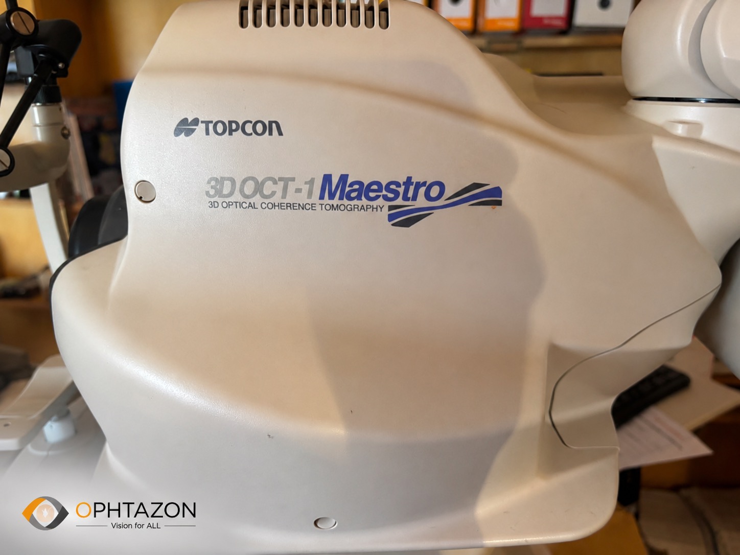
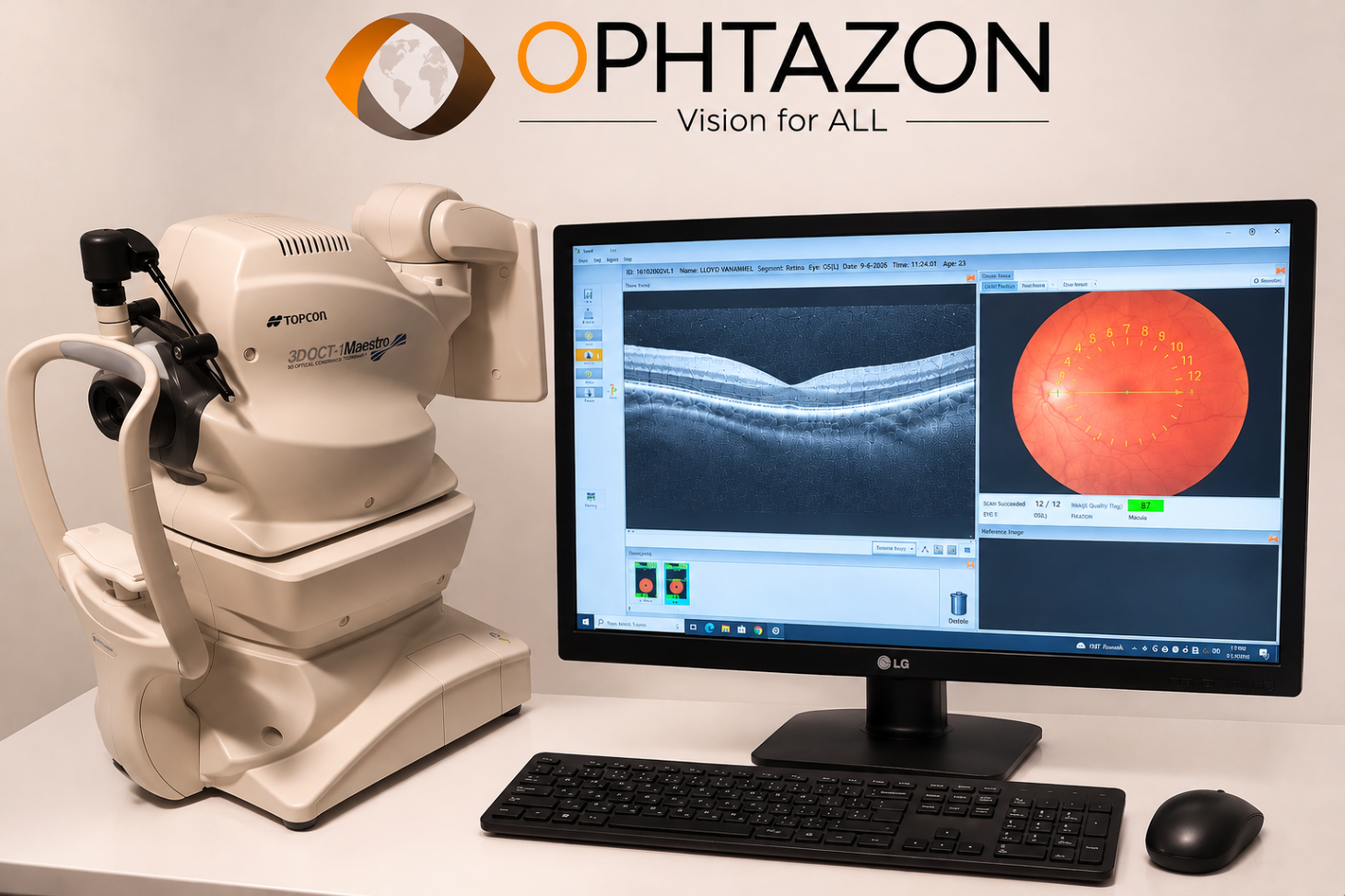
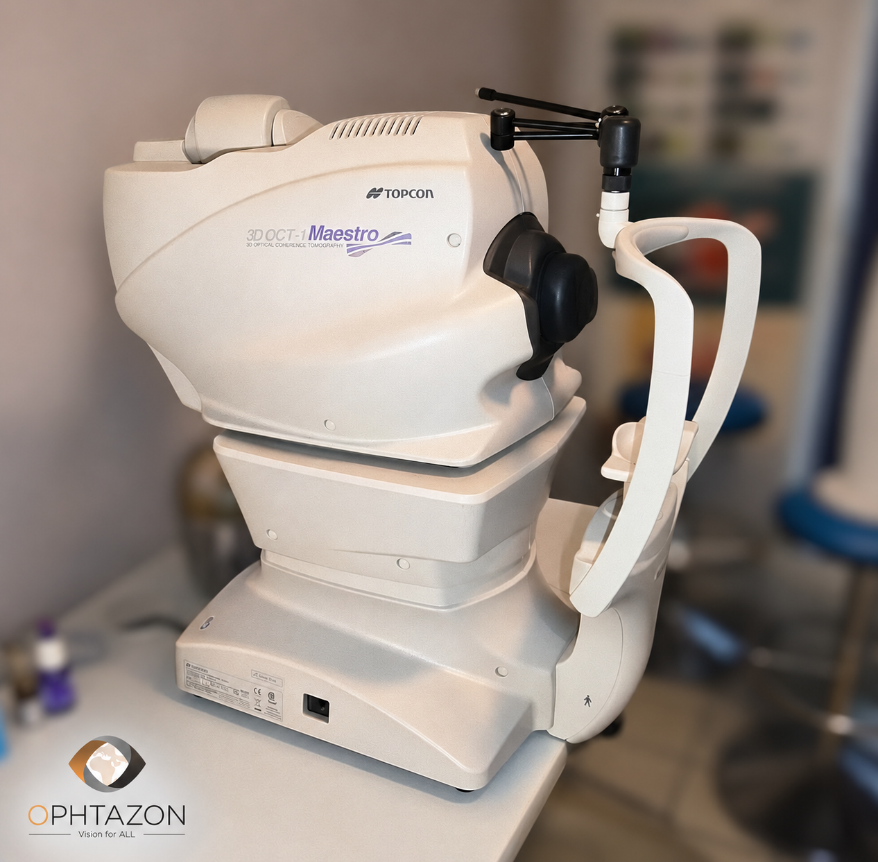
TOPCON 3D OCT-1 Maestro (Maestro I)
The TOPCON 3D OCT-1 Maestro, also known as the Maestro I, is an ophthalmic imaging device that combines spectral OCT (SD-OCT) with a non-mydriatic color retinograph.
It enables examinations of the retina, macula, and optic nerve head, with automated image acquisition that simplifies its use in an ophthalmology practice.
Technical Specifications
Manufacturer: TOPCON
Model: 3D OCT-1 Maestro
Technology: Spectral OCT (SD-OCT)
Acquisition speed: 50,000 A-scans/second
Axial resolution: Approximately 6 µm in tissue
OCT wavelength: Approximately 840 nm
Retinography: Non-mydriatic color
Field of view: 45°
Acquisition: Automatic, with alignment and autofocus
Examinations: Macula, optic disc, RNFL, ganglion cells
OCT angiography: Not available in standard configuration
Interface: Integrated touchscreen
Main Clinical Applications
- Macula: analysis of retinal layers, AMD, macular edema, and epiretinal membranes.
- Glaucoma: measurement of retinal nerve fiber layer (RNFL) thickness, optic disc analysis, and ganglion cells.
- Retinography: Color fundus photography.
- Patient follow-up: Comparison of examinations and analysis of disease progression.
Key Features
- OCT acquisition and color retinography in a single session.
- 12 × 9 mm wide-field scan covering the macula and optic disc.
- Fully automated operation.
- Compact design, weighing approximately 21 kg.
- Anterior segment imaging possible with the optional HA-2 adapter.

Checked by OPHTAZON
 1
Year of mfg.
2015
Available on 2027-01-20

Checked by OPHTAZON
 3
Year of mfg.
2018
Available on 2027-01-21
Checked by OPHTAZON
 2
Year of mfg.
2010
Available on 2026-12-28
There was an error when considering the interest of the product. Please try or contact the support.

Your partnership request has been sent by email to the OPHTAZON team.
Your partnership request has been sent by email to the OPHTAZON team.
Your application for a partnership was not successful. Please try again or contact the support.
HOME
sales@ophtazon.com / +33 141 515 490
INTERNATIONAL SALES MANAGER
M. Sadreddine MEHRI
sadreddine.mehri@ophtazon.com / +33 6 08 84 07 42
TECHNICAL & SALES MANAGER IVORY COAST
M. Khaled SELLAMI
khaled.sellami@ophtazon.com / +225 07 68 46 36 14
Nous utilisons différentes technologies pour optimiser votre expérience utilisateur.
Merci de cliquer sur le bouton pour donner votre accord.
Vous souhaitez en savoir plus ? Cliquez ici

 Ophtazon
 Ophtazon Ivory Coast
 Ophtazon Sénégal
 Ophtazon Cameroon
 Ophtazon Togo

